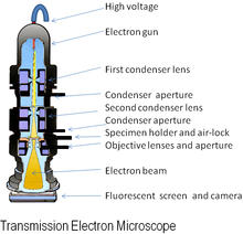
Tiān-chú hián-bî-kiàⁿ
Tēn-tsú hén-bî-kiàñ (ing-gí: Electron microscope; kán-tshing: tēn kiañ) sī sú-iōng tēn-tsú lâi hén-sī bu̍t-kiāñ ê lāi-pōo hi̍k-tsiá piáu-bīn ê hén-bî-kiàñ. Ko-sok ê tēn-tsú ê pho-tn̂g pí khó-kiàn kng ê pho-tn̂g khah té (pho-lia̍p jī-siōng-sìng), lî-tshiáñ tēn-tsú hén-bî-kiàñ ê hun-pēn-lu̍t (tāi-iok 0.2 nāi-bí) pí kng-ha̍k hén-bî-kiàñ ê hun-pēn-lu̍t (tāi-iok 200 nāi-bí) koh kha kuân tsiâñ-tsē.[1]


Ìng-iōng
siu-kái- Puàn-tō-thé hām tsu-liāu thú-tsûn
- Circuit edit[2]
- Defect analysis[3]
- Kòo-tsiong hun-sik[4]
Sing-bu̍t-ha̍k hām sèñ-miā kho-ha̍k
- Kē-un sing-bu̍t-ha̍k[5]
- Cryo-electron microscopy[6]
- Diagnostic electron microscopy[7]
- Drug research (e.g. antibiotics)[8]
- Electron tomography[9]
- Lia̍p-tsú hun-sik[10]
- Particle detection[11]
- Protein localization[12]
- Kiat-kòo sing-bu̍t-ha̍k[6]
- Tissue imaging[13]
- Toxicology[14]
- Pēñ-to̍k-ha̍k (e.g. pēñ-to̍k tsài-liōng monitoring)[15]
- Tsâi-liāu gén-kiú
- Device testing and characterization[16]
- Tōng-thài tsâi-liāu si̍t-giām[17]
- Electron beam-induced deposition[18]
- In-situ characterisation[19]
- Materials qualification[20]
- Medical research[8]
- Nāi-bí kè-liōng-ha̍k[21]
- Nanoprototyping[22]
- Kok-tsíng hâng-gia̍p
- Chemical/Petrochemical[23]
- Direct beam-writing fabrication[24]i-iáu
- Food science[25]
- Huat-i-ha̍k[26]
- Kim-sio̍k tuàn-bīn hén-bî-kiàñ kuan-tshat-huat (Tuàn-khió siòng-ha̍k)[27]
- Micro-characterization[28]
- Mining (khòng-bu̍t sik-hòng hun-sik)[29]
- I-iáu phín-khòng (Pharmaceutical QC)[30]
Tsù-kái
siu-kái- ↑ Erni, Rolf; Rossell, MD; Kisielowski, C; Dahmen, U (2009). "Atomic-Resolution Imaging with a Sub-50-pm Electron Probe". Physical Review Letters. 102 (9): 096101. Bibcode:2009PhRvL.102i6101E. doi:10.1103/PhysRevLett.102.096101. PMID 19392535. (Eng-gí)
- ↑ Boehme, L.; Bresin, M.; Botman, A.; Ranney, J.; Hastings, J.T. (2015). "Focused electron beam induced etching of copper in sulfuric acid solutions". Nanotechnology. 26 (49): 495301. Bibcode:2015Nanot..26W5301B. doi:10.1088/0957-4484/26/49/495301. PMID 26567988.
- ↑ Kacher, J.; Cui, B.; Robertson, I.M. (2015). "In situ and tomographic characterization of damage and dislocation processes in irradiated metallic alloys by transmission electron microscopy". Journal of Materials Research. 30 (9): 1202–1213. Bibcode:2015JMatR..30.1202K. doi:10.1557/jmr.2015.14.
- ↑ Rai, R.S.; Subramanian, S. (2009). "Role of transmission electron microscopy in the semiconductor industry for process development and failure analysis". Progress in Crystal Growth and Characterization of Materials. 55 (3–4): 63–97. doi:10.1016/j.pcrysgrow.2009.09.002.
- ↑ Morris, G.J.; Goodrich, M.; Acton, E.; Fonseca, F. (2006). "The high viscosity encountered during freezing in glycerol solutions: Effects on cryopreservation". Cryobiology. 52 (3): 323–334. doi:10.1016/j.cryobiol.2006.01.003. PMID 16499898.
- ↑ 6.0 6.1 von Appen, Alexander; Beck, Martin (May 2016). "Structure Determination of the Nuclear Pore Complex with Three-Dimensional Cryo electron Microscopy". Journal of Molecular Biology. 428 (10): 2001–10. doi:10.1016/j.jmb.2016.01.004. PMC 4898182 . PMID 26791760.
- ↑ Florian, P.E.; Rouillé, Y.; Ruta, S.; Nichita, N.; Roseanu, A. (2016). "Recent advances in human viruses imaging studies". Journal of Basic Microbiology. 56 (6): 591–607. doi:10.1002/jobm.201500575. PMID 27059598.
- ↑ 8.0 8.1 Cushnie, T.P.; O’Driscoll, N.H.; Lamb, A.J. (2016). "Morphological and ultrastructural changes in bacterial cells as an indicator of antibacterial mechanism of action". Cellular and Molecular Life Sciences. 73 (23): 4471–4492. doi:10.1007/s00018-016-2302-2. PMID 27392605.
- ↑ Li, M.-H.; Yang, Y.-Q.; Huang, B.; Luo, X.; Zhang, W.; Han, M.; Ru, J.-G. (2014). "Development of advanced electron tomography in materials science based on TEM and STEM". Transactions of Nonferrous Metals Society of China. 24 (10): 3031–3050. doi:10.1016/S1003-6326(14)63441-5.
- ↑ Li, W.J.; Shao, L.Y.; Zhang, D.Z.; Ro, C.U.; Hu, M.; Bi, X.H.; Geng, H.; Matsuki, A.; Niu, H.Y.; Chen, J.M. (2016). "A review of single aerosol particle studies in the atmosphere of East Asia: morphology, mixing state, source, and heterogeneous reactions". Journal of Cleaner Production. 112 (2): 1330–1349. doi:10.1016/j.jclepro.2015.04.050.
- ↑ Sousa, R.G.; Esteves, T.; Rocha, S.; Figueiredo, F.; Quelhas, P.; Silva, L.M. (2015). Automatic detection of immunogold particles from electron microscopy images. Image Analysis and Recognition. Lecture Notes in Computer Science. 9164. pp. 377–384. doi:10.1007/978-3-319-20801-5_41. ISBN 978-3-319-20800-8.
- ↑ Perkins, G.A. (2014). "The Use of miniSOG in the Localization of Mitochondrial Proteins". Mitochondrial Function. Methods in Enzymology. 547. pp. 165–179. doi:10.1016/B978-0-12-801415-8.00010-2. ISBN 9780128014158. PMID 25416358.
- ↑ Chen, X.D.; Ren, L.Q.; Zheng, B.; Liu, H. (2013). "Physics and engineering aspects of cell and tissue imaging systems: microscopic devices and computer assisted diagnosis". Biophotonics in Pathology: Pathology at the Crossroads. 185 (Biophotonics in Pathology): 1–22. doi:10.3233/978-1-61499-234-9-1. PMID 23542929.
- ↑ Fagerland, J.A.; Wall, H.G.; Pandher, K.; LeRoy, B.E.; Gagne, G.D. (2012). "Ultrastructural analysis in preclinical safety evaluation". Toxicologic Pathology. 40 (2): 391–402. doi:10.1177/0192623311430239. PMID 22215513.
- ↑ Heider, S.; Metzner, C. (2014). "Quantitative real-time single particle analysis of virions". Virology. 462–463: 199–206. doi:10.1016/j.virol.2014.06.005. PMC 4139191 . PMID 24999044.
- ↑ Tsekouras, G.; Mozer, A.J.; Wallace, G.G. (2008). "Enhanced performance of dye sensitized solar cells utilizing platinum electrodeposit counter electrodes". Journal of the Electrochemical Society. 155 (7): K124–K128. Bibcode:2008JElS..155K.124T. doi:10.1149/1.2919107.
- ↑ Besenius, P.; Portale, G.; Bomans, P.H.H.; Janssen, H.M.; Palmans, A.R.A.; Meijer, E.W. (2010). "Controlling the growth and shape of chiral supramolecular polymers in water". Proceedings of the National Academy of Sciences of the United States of America. 107 (42): 17888–17893. Bibcode:2010PNAS..10717888B. doi:10.1073/pnas.1009592107. PMC 2964246 . PMID 20921365.
- ↑ Furuya, K. (2008). "Nanofabrication by advanced electron microscopy using intense and focused beam". Science and Technology of Advanced Materials. 9 (1): Article 014110. Bibcode:2008STAdM...9a4110F. doi:10.1088/1468-6996/9/1/014110. PMC 5099805 . PMID 27877936.
- ↑ Kosasih, Felix Utama; Ducati, Caterina (May 2018). "Characterising degradation of perovskite solar cells through in-situ and operando electron microscopy". Nano Energy. 47: 243–256. doi:10.1016/j.nanoen.2018.02.055.
- ↑ Maloy, Stuart A.; Sommer, Walter F.; James, Michael R.; Romero, Tobias J.; Lopez, Manuel R.; Zimmermann, Eugene; Ledbetter, James M. (13 May 2017). "The Accelerator Production of Tritium Materials Test Program". Nuclear Technology. 132 (1): 103–114. doi:10.13182/nt00-a3132.
- ↑ Ukraintsev, Vladimir (27 February 2012). "Review of reference metrology for nanotechnology: significance, challenges, and solutions". Journal of Micro/Nanolithography, MEMS, and MOEMS. 11 (1): 011010. doi:10.1117/1.JMM.11.1.011010.
- ↑ Wilhelmi, O.; Roussel, L.; Faber, P.; Reyntjens, S.; Daniel, G. (June 2010). "Focussed ion beam fabrication of large and complex nanopatterns". Journal of Experimental Nanoscience. 5 (3): 244–250. Bibcode:2010JENan...5..244W. doi:10.1080/17458080903487448.
- ↑ Vogt, E.T.C.; Whiting, G.T.; Chowdhury, A.D.; Weckhuysen, B.M. (2015). Zeolites and zeotypes for oil and gas conversion. Advances in Catalysis. 58. pp. 143–314. doi:10.1016/bs.acat.2015.10.001. ISBN 9780128021262.
- ↑ Lai, Shih-En; Hong, Ying-Jhan; Chen, Yu-Ting; Kang, Yu-Ting; Chang, Pin; Yew, Tri-Rung (18 September 2015). "Direct-Writing of Cu Nano-Patterns with an Electron Beam". Microscopy and Microanalysis. 21 (6): 1639–43. Bibcode:2015MiMic..21.1639L. doi:10.1017/S1431927615015111. PMID 26381450.
- ↑ Sicignano, Angelo; Di Monaco, Rossella; Masi, Paolo; Cavella, Silvana (October 2015). "From raw material to dish: pasta quality step by step". Journal of the Science of Food and Agriculture. 95 (13): 2579–87. doi:10.1002/jsfa.7176. PMID 25783568.
- ↑ Brożek-Mucha, Zuzanna (2014). "Scanning Electron Microscopy and X-Ray Microanalysis for Chemical and Morphological Characterisation of the Inorganic Component of Gunshot Residue: Selected Problems". BioMed Research International. 2014: 428038. doi:10.1155/2014/428038. PMC 4082842 . PMID 25025050.
- ↑ Carbonell-Verdu, A.; Garcia-Sanoguera, D.; Jorda-Vilaplana, A.; Sanchez-Nacher, L.; Balart, R. (2016). "A new biobased plasticizer for poly(vinyl chloride) based on epoxidized cottonseed oil". Journal of Applied Polymer Science. 33 (27): 43642.
- ↑ Ding, Jie; Zhang, Zhiming (1 May 2015). "Micro-characterization of dissimilar metal weld joint for connecting pipe-nozzle to safe-end in generation III nuclear power plant". Acta Metall Sin. 51 (4): 425–39. doi:10.11900/0412.1961.2014.00299 (inactive 28 February 2022).
- ↑ Tsikouras, Basilios; Pe-Piper, Georgia; Piper, David J.W.; Schaffer, Michael (June 2011). "Varietal heavy mineral analysis of sediment provenance, Lower Cretaceous Scotian Basin, eastern Canada". Sedimentary Geology. 237 (3–4): 150–165. Bibcode:2011SedG..237..150T. doi:10.1016/j.sedgeo.2011.02.011.
- ↑ Li, Xiang; Jiang, Chuan; Pan, Lili; Zhang, Haoyang; Hu, Lang; Li, Tianxue; Yang, Xinghao (15 July 2014). "Effects of preparing techniques and aging on dissolution behavior of the solid dispersions of NF/Soluplus/Kollidon SR: identification and classification by a combined analysis by FT-IR spectroscopy and computational approaches". Drug Development and Industrial Pharmacy. 41 (1): 2–14. doi:10.3109/03639045.2014.938080. PMID 25026247.
Tsham-ua̍t
siu-kái- Thiò-siā tiān-chú hián-bî-kiàⁿ (Thiò-siā tēn-chú hén-bî-kiàñ/Transmission electron microscopy)
- Hah-hì (Yawn)
- Bí-kok sòo Cruikshank àn (United States v. Cruikshank)
- Bí-kok sòo Miller àn (United States v. Miller)*Discord
- San-tiok (san-tik; mangosteen; purple mangosteen)
Guā-pōo lên-ket
siu-kái- An Introduction to Electron Microscopy Archived 2013-07-19 at the Wayback Machine.: resources for teachers and students
- Cell Centered Database – Electron microscopy data
- Science Aid: Electron Microscopy Archived 2009-07-30 at the Wayback Machine.: high school (GCSE, A Level) resource
It-puañ
siu-kái- Animations and explanations of various types of microscopy including electron microscopy (Université Paris Sud)
- Environmental Scanning Electron Microscopy (ESEM)
- ETH Zurich website: graphics and images illustrating various procedures
- Eva Nogales's Seminar: "Introduction to Electron Microscopy" Archived 2022-02-15 at the Wayback Machine.
- FEI Image Contest: FEI has had a microscopy image contest every year since 2008
- Introduction to electron microscopy by David Szondy
- Nanohedron.com image gallery: images generated by electron microscopy
- X-ray element analysis in electron microscopy: information portal with X-ray microanalysis and EDX contents
Li̍k-sú
siu-kái- John H.L. Watson's recollections at the University of Toronto when he worked with Hillier and Prebus Archived 2019-03-30 at the Wayback Machine.
- Rubin Borasky Electron Microscopy Collection, 1930–1988 (Archives Center, National Museum of American History, Smithsonian Institution)